Zooplankton Under the Microscope
 No comments
 As you may have seen in one of our earlier dispatches regarding the zooplankton sampling methods (to read the dispatch click here) the zooplankton team scours the ocean with two nets, the smaller of which samples to a depth of 300 meters, nearly 1,000 feet below the surface (think of the Eiffel Tower standing between the net and the surface.) While the zooplankton group focuses on Antarctic krill (Euphausia superba, the largest and most abundant of the many species of crustaceans collectively called “krill,” a word that means whale food in Norwegian,) there are many other zooplankters we come across that also play an important role in the Southern Ocean ecosystem. One of the many strengths of the LTER program is the ability to look at long term changes in abundance (number and/or biomass) of these different species and the regional community composition (the relative amounts of each species.)
The LTER’s sampling grid covers inshore and offshore waters along the Antarctic Peninsula, where we see a broad range of organisms including jellyfish, squid, fish, salps, worms, and amphipods, among others.
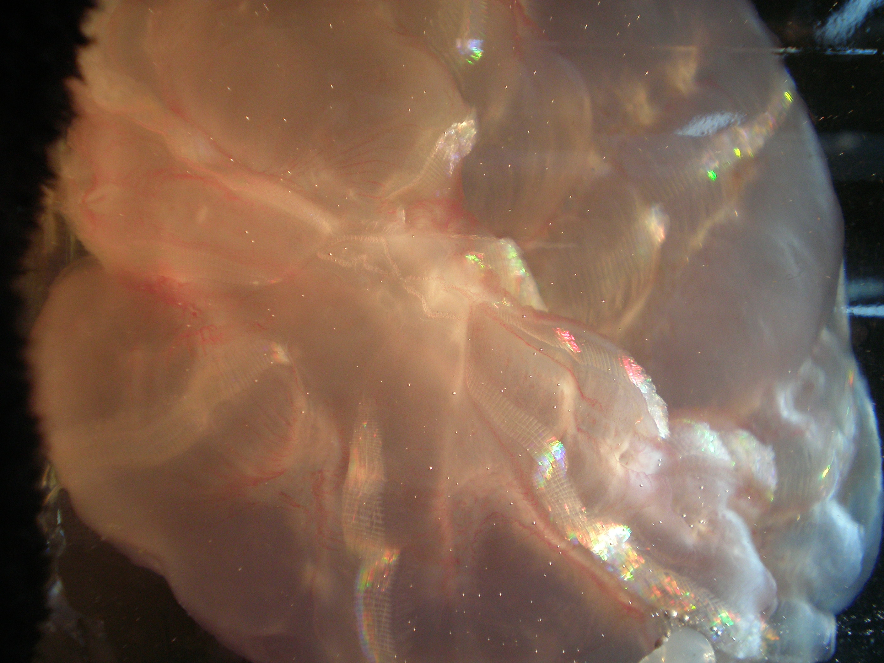
A ctenophore: a creature similar to jellyfish but without any stinging cells. These populate cold waters in both Antarctica and the Arctic.
One of our dissecting microscopes, used in the analysis of these organisms, is equipped with a digital camera that allows us to take pictures of what we see. We decided to compile some of our favorites here with the magnification for scale.
In the upper left is a pteropod called Limacina. Pteropods (a name meaning winged foot) are similar to snails and slugs, only they have wings and swim around in the ocean. Limacina swim with a bat-like flapping motion of their wings (the “chinchilla ears” in the photo). Also of note is their mucus net used in feeding; a single Limacina, roughly the size of the “G” on your keyboard, can cast a mucus net many times its size in diameter. This net traps phytoplankton until the Limacina consumes the net: algae, mucus and all. This architectural feat was discovered by blue-water divers watching pteropods feed in the ocean far from land.
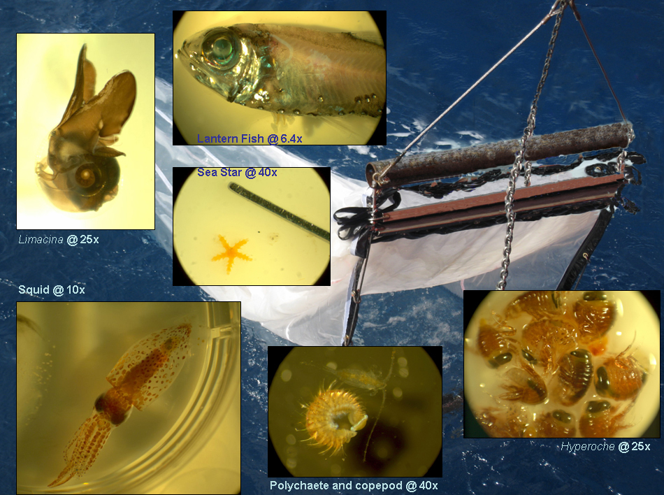
These closeups, taken through a microscope, are layered against a picture of the net tow being brought back aboard the ship.
At the top-center of the picture is a species of Protomyctophum, or Lantern Fish. The pearl-like spheres along its body are called photophores. Photophores are light-producing organs that typically line the bottom of a fish (though not only in fish, Euphausia superba has them too) in order to break up its silhouette, making it difficult for predators to identify them as prey.
In the middle of the picture is a very, very small starfish. To give you an idea of how small, the width of the forceps next to it is roughly 0.25 millimeters! This interesting pelagic (which is strange for the mostly bottom dwelling starfish) creature has shown up in two catches, hundreds of kilometers apart. None of us know exactly who or what it is.
The three pictures on the bottom are of a squid, a polychaete worm curled up next to a copepod (a small planktonic crustacean) and a group of Hyperoche amphipods. Amphipods are another group of crustaceans. This particular species is one of the more common amphipods found in the study. Note the large eyes! The squid and the polychaete, on the other hand, are rare finds. These photos offer a glimpse of the high diversity of small animals living in the Southern Ocean.